Optic Atrophy Diagnosis and Prevention
#LIVE2.0 #Review
Poor night vision and blurred vision are just some of the symptoms experienced by people with optic nerve damage caused by optic atrophy, an eye condition that affects the optic nerve, which connects the eye to the brain.
The optic nerve is made up of two nerve fibers, one of which carries visual information from the eye to the brain and the other of which carries visual information from the brain to the eye.
Click here if you’re interested in learning more about the symptoms of optic atrophy.
To diagnose optic atrophy, it is necessary for an individual to be thoroughly examined by a physician.
How Is Optic Atrophy Diagnosed?
Optic atrophy is a diagnostic label when the optic nerve is visibly smaller than in normal eyes. This condition can be caused by a variety of conditions, so it is important to determine what has caused the small optic nerve.
The patient’s history and visual symptoms provide clues to the cause, but there are no definitive tests at this time that can diagnose the underlying cause based upon symptoms. As a result, several eye examinations are performed to learn more about the eye and how it works.

Imaging techniques, such as MRI or CT scan, may be performed. These tests can help determine whether a disease of another organ may be causing damage to the optic nerve.
The most common way of diagnosing this condition is through an MRI (magnetic resonance imaging) scan of the head because it can detect damage in the optic nerve caused by inflammation, glaucoma, or other diseases.
The MRI helps doctors determine whether there are any abnormalities in the brain or spinal cord.
An MRI of the head may be performed on a patient who has experienced a traumatic injury or stroke, as well as on patients with other conditions such as multiple sclerosis (MS), multiple system atrophies (MSAs), and Parkinson’s disease. In addition to being used as a diagnostic tool, an MRI can be used to monitor a patient’s progress while receiving treatment for optic atrophy.
What Eye Examinations Are Used to Diagnose Optic Atrophy?
Several examinations and tests are performed to detect symptoms and potential eye injuries that may have caused optic atrophy.
The first part of the exam will include reading letters on an eye chart.
A visual acuity test determines how well a person can see across a range of distances. The person being tested will cover one eye at a time and read aloud, beginning with the smallest line of letters they can see. This will be repeated for each eye individually.
If neither eye has a problem, the doctor will repeat the test with both eyes open to check if the problem is caused by poor vision or poor eye coordination. The doctor may detect a binocular condition such as strabismus if using both eyes causes visual acuity to decline.
This test may be repeated again after dilating (widening) the pupils to see if there are any hidden signs of optic nerve damage in addition to those already observed.
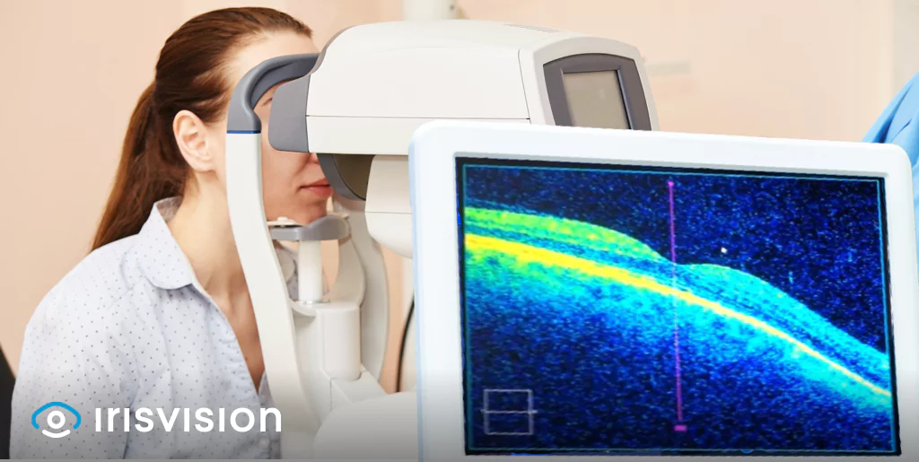
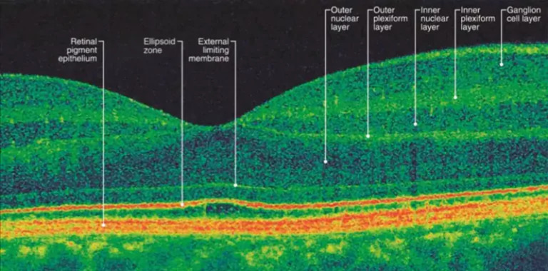
Other tests include Optical coherence tomography (OCT), which is a non-invasive imaging test that images the retina (the light sensitive tissue at the back of the eye) and optic nerve. It provides information about changes in retinal structure and thickness, as well as changes in the size of the optic nerve head and cup size.
Source: American Academy of Ophthalmology
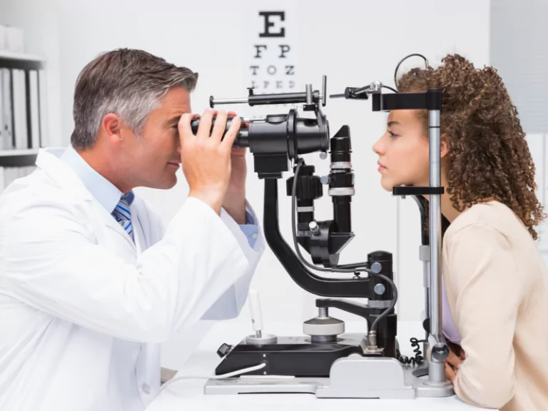
Your doctor may also opt for a Slit lamp examination, a process where a special microscope called a slit lamp is used to evaluate structures at the front of your eyes in great detail, including eyelids, lashes, cornea, iris, lens and examine your eyes for signs of a disease or an eye injury.
After applying numbing drops to your eyes, your doctor shines a slit lamp into your eyes from different angles to closely examine the tissue of your eyes.
The eye specialist may also dilate your pupils so that they can examine the optic nerves in the back of each eye. The eye doctor will also look at your optic disc, which is where your optic nerve enters the back of your eye.
If you have optic atrophy, the color and shape of your optic nerve might be abnormal. Your doctor might also see a white spot called pallor on your optic disc. These symptoms are all signs that you have optic atrophy.
Another way to examine the symptoms of optic atrophy is through Ophthalmoscopy. An instrument called an ophthalmoscope is used, which allows an eye doctor to examine the retina, optic nerve, and other structures inside your eyes. This instrument allows the physician to see the back of the eye, including the optic nerve.
For special cases and eye conditions such as glaucoma, which can severely damage the optic nerve, tonometry is a test that measures the pressure inside your eyes (intraocular pressure).
Tips to Prevent Optic Nerve Damage and Prevent Vision Loss
Vision loss can be caused by a variety of eye conditions and diseases, although many of them have similar preventative techniques. Let’s look at some of the most critical but simple preventive measures you may take to lower your risk of eye issues and protect your optic nerve from damage.
Be aware of medications that may increase pressure inside the eye (intraocular pressure). Some drugs used to treat high blood pressure can increase intraocular pressure. Talk with your doctor about these risks and benefits when taking any new medication.
Use eye drops exactly as prescribed. If you stop using them, pressure inside the eye will rise, resulting in continued optic nerve damage. Taking too few drops can reduce their effectiveness and lead to higher pressure inside the eye. If eye drops are not enough to control your glaucoma, discuss other treatments with your eye doctor.
Exercising regularly and maintaining a healthy weight will help keep your blood pressure under control and prevent additional effects on the optic nerve.
Ensure that your children receive regular eye exams in order to detect any early warning signs or symptoms of eye conditions.
Use sunglasses with UV protection while going out in the sun, as UV rays are harmful to the eyes.
People who wear contact lenses should follow proper guidelines for lens cleaning and wear them only after consulting an eye specialist.
Don’t let your eyes get red or itchy because of dust allergies or pollen allergies, as this can result in glaucoma, which is a major cause of optic nerve damage.
If you experience any discomfort in your eyes, see an ophthalmologist right away.
Other tips include:
-
Avoid smoking as it increases your risk of optic nerve damage and other eye conditions
-
Control chronic health conditions such as diabetes or another chronic health condition, working with your doctor to control them
-
Know the risk factors for optic nerve damage
-
Keep your blood pressure under control
-
Eating a healthy diet
-
Protecting your eyes from injury
Optic atrophy can’t be cured, but it can be treated with medication in some cases. People with thyroid disease should also manage their condition effectively to reduce their risk of optic atrophy due to a pituitary tumor or thyroid eye disease.
Assistive technology like IrisVision is ideal for people with low vision caused by optic atrophy. These wearable low vision aids leverage award-winning software lenses to help users magnify images at 14X and regain their lost peripheral vision by providing an industry leading 77 field of view.
That’s not all. With IrisVision, you can stream videos on YouTube, capture and store images, as well as stay connected to the outside world with social media. Click here for a free 30-day trial session.