Have you experienced floaters in your vision or a black curtain falling over your field of vision? If so, you have likely experienced a retinal tear or small retinal detachment. Leaving it unattended for too long will lead to legal blindness. Taking the necessary steps before it gets out of hand is necessary.
Are you among the ones who have already taken the steps for retinal reattachment? Or are you among those who are soon to undergo retinal detachment surgery?
Either way, you are in the right place. We are here to help you understand what happens during and after retinal detachment surgeries.
Let’s recap a little on the types of retinal detachment.
It’s known that there are mainly three types of retinal detachments:Rhegmatogenous, Traction, and Exudative. Each has varying causes of retinal detachment, but Rhegmatogenous retinal detachment is the most common type. Click here to learn more about the types of retinal detachment.
What Does Retinal Detachment Do to Eyesight?
As the retina senses light and projects images to the brain to see properly, if the retina is detached from its original position, it won’t be able to perform its necessary duties. Hence, vision becomes blurry, there is a sudden appearance of floaters and light flashes, and vision is obscured. Based on the severity and location of the retinal tear, the central or peripheral vision is affected on a greater scale.
Are There Several Procedures for Retinal Tears and Detachments?
Retinal detachment is a sudden eye condition. It happens without any warning and shouldn’t be taken too lightly. Age is the most common cause, yet people are prone to developing retinal detachment if an existing retinal tear is present or due to head injuries.
To avoid developing a retinal detachment, an ophthalmologist may perform one or two noninvasive procedures to repair the tear and seal the retina to the back of the eye. These procedures are only done when the retinal detachment is not too severe.
Most retinal tears are treated with laser photocoagulation and cryotherapy. Both retinal detachment procedures are used to prevent the tear or detachment from growing bigger.
Laser Photocoagulation
Laser photocoagulation focuses on the retinal tear or small detachment. The laser burns the area around the retinal tear or detachment to create scar tissue. This helps seal the tear or reattach the detached portion of the retina to the underlying tissue.
Through this procedure, the fluid, i.e., vitreous, is prevented from traveling under the retina and stops the detachment.
Cryotherapy
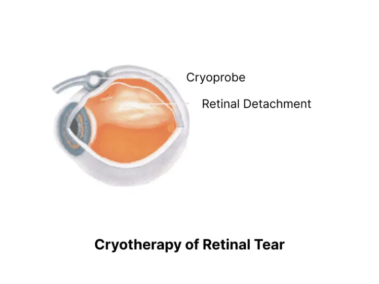
A freezing therapy, where the cold is used to create scar tissue in the retina. A freezing probe is placed over the tear or small detached area. Every time a specific area is frozen, scar tissues are formed that help seal the tear or reattach the retina to the underlying tissues in its correct position.
Retinal Detachment Surgery
Retinal detachment surgeries are performed when the detachment is too big to be treated through laser photocoagulation or cryotherapy procedures. There are mainly three types of retinal detachment surgeries:
-
Pneumatic Retinopexy
-
Scleral Buckle
-
Vitrectomy
The type of retinal detachment surgery is determined based on different factors, like location and size of the detachment, and whether the person has had prior cataract surgery.
Pneumatic Retinopexy
During this surgery for retinal detachment, a small amount of the fluid, called vitreous, is removed. Following that, a small amount of expanding gas is injected to push the retina back to its place at the back of the eye. In some cases, to secure the retina in its original position, cryotherapy may be used.
The following precautions must be taken post retinal detachment surgery:
-
Keep the head in a specific position for the bubble to stay in the right place.
-
Avoid air travel, intense exercise, and heavy lifting.
Scleral Buckle
Sclera is the white part of the eye. During this surgery, a small, flexible band is placed around the sclera. This tiny band puts gentle pressure on the sides of the eye and slowly pushes it inside towards the retina. Therefore, it helps the retina reattach to its original position.
The following should be adhered to after the detached retina surgery:
-
Wear an eye patch or bandage over the eyes for a few days.
-
Avoid heavy lifting and intense exercise to prevent putting additional pressure on the eyes.
Vitrectomy
During this surgery for detached retina, a small cut is made in the sclera, through which a small quantity of vitreous is sucked out. Other procedures, such as laser photocoagulation or cryotherapy, may be used to treat or repair any tears or scars on the retina. Additionally, a gas bubble might be used to hold the retina in its original position.
Ensure the following after detached retina surgery:
-
Use antibiotic eye drops to avoid infection.
-
Avoid air travel, heavy lifting, and intense exercise.
What to Expect After Retinal Detachment Surgery?
As soon as the retinal detachment surgery is completed, the treated eye is covered with some protective shields or patches. In most cases, the patients are sent home on the same day, but based on the doctor’s discretion, they might be asked to stay for a day or two.
Let’s talk about the real thing now: what to expect…
During the retinal detachment recovery time:
-
Slight discomfort is felt in the eyes, especially in scleral buckle surgery.
-
Vision becomes blurry for a few weeks.
-
Avoid rubbing your eyes and follow all the precautions.

It’s expected that the vision will return to normal in about 6 weeks. Yet, in the case of a detached macula, the sight may not be as clear as before.
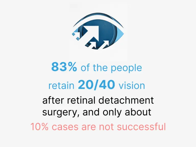
The outlook of eyesight after retinal detachment surgery depends on the type, severity, and whether the macula has been detached or not. If the macula stays attached, it’s suggested that 83% of the people retain 20/40 vision after retinal detachment surgery, and only about 10% of the cases cases are not successful in retinal reattachment.
Retinal Detachment Recovery Time
Under normal circumstances, it takes between two and four weeks to recover from retinal detachment surgery. Yet, it depends on the eye condition and how well it is taken care of after retinal detachment surgery.
Proper recovery time must be ensured and some of the following should be kept in mind:
-
For those who have been treated with a gas bubble, proper face down positioning is important. Use a face down apparatus to help with the face positioning.
-
Use postoperative drops to avoid inflammation and bacterial infections.
-
Limit your activities and avoid bending or heavy lifting.
-
Wear protective shields for at least a week and follow guidelines to ensure the incision heals properly after retinal detachment surgery.
-
Avoid flying, as the gas bubble could expand, which would lead to more serious problems.
-
Always wear the green bracelet till the gas dissipates completely.
-
Most importantly, be patient.
Does that clear up any concerns related to retinal detachment surgery and recovery? We have another question that comes to mind when thinking about the 8 to 10% of unsuccessful restorative attempts. What do you do if you are part of that 8 to 10%? Is there a way to retain or enhance eyesight after retinal detachment surgery?
The best solution is to use low-vision aids. We’ll be further discussing the steps that can be taken with the help of low vision aids post retinal detachment surgery.
Low Vision Aids for Vision Post Retinal Detachment Surgery
Low vision aids are prescribed when the vision can’t be corrected through glasses, contact lenses, or surgery. Normally, retinal detachment treatments are highly effective and have a 90% success rate, yet there are still chances that vision after retinal detachment surgery is not fully restored. In such cases, glasses or contact lenses are of no help and require visual aids for retinal detachment to assist in retaining the leftover vision. Some visual aids used are magnifiers, standing and hand-held magnifiers, strong magnifying reading glasses, loupes, and small telescopes.
In his article “Patient Use of Low-Vision Aids after Retinal Detachment Surgery”, I M Chan stated:
“Patient use of low vision aids to improve postoperative vision was evaluated in 32 eyes after successful repair of a retinal detachment that involved the macular area. Despite best-corrected postoperative visual acuity ranging from 5/155 to 10/38, both distance and near vision could be improved in all cases using telescopic and microscopic systems, respectively. Patient acceptance of the telescopic systems was 62%, and of the microscopic systems, 96%.” (I M Chan, 1985).
IrisVision provides visual aids for retinal detachment that use award-winning assistive technology to make use of the remaining vision. Our assistive device, IrisVision Vista, is a wearable, hands-free low-vision headset controlled by voice or a simple remote.
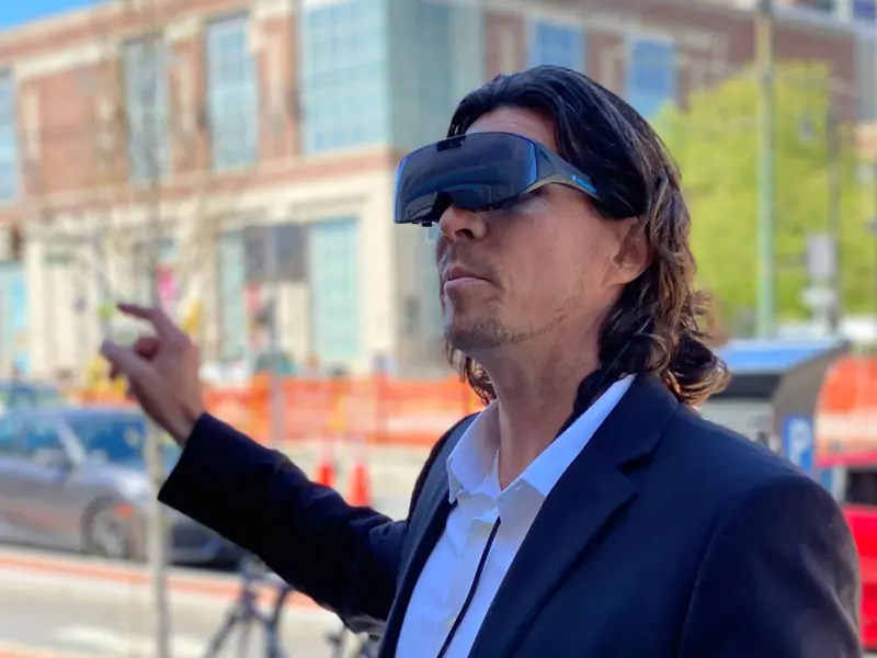
It’s an easy-to-use system that enables people who have gone through retinal detachment surgery and have experienced impaired vision to use it.
Vision after retinal detachment surgery is usually not affected. However, if the retina has been detached for a long time, especially the macula, only some vision will return and it will be less than 20/200, which is legally blind. Even after months of retinal detachment recovery, the vision may not fully return.
As a visual aid for retinal detachment, IrisVision Vista is among the most effective devices and is endorsed by the Innovation Tri-Valley Leadership Group’s #GameChangers Award.

IrisVision Vista employs advanced technology that helps people with low vision navigate modes according to the requirements and low vision condition of a person.
Vista is equipped with a wide range of vision modes, each designed to target a certain low vision condition faced after retinal detachment surgery.
Scene: It provides a 70-degree field of view and 14X magnification, allowing the user to view at greater distances and focus on a certain object. This mode is best when central vision is affected. It utilizes the remaining vision and magnifies it for the user to view a high-definition view. People who have suffered macula detachment can benefit from it for their vision after retinal detachment surgery has been affected.
Retinitis Pigmentosa: The RP mode is exclusively designed for people suffering from retinitis pigmentosa. However, any peripheral vision loss can be retained with this mode, even after retinal detachment surgery. The software lens helps the user change the field of vision, adjusting it to fit the whole scene within the visual frame. All field restrictions are removed to augment the remaining vision for different viewing tasks.
These two modes address the peripheral and central vision loss that may be experienced after detached retina surgery. However, there are other features that help users view and enjoy different aspects of everyday life.
Iris: The voice-first Iris assistant lets you skim through a textbook with your eyes shut. It uses on-device OCR (optical character recognition) technology to scan text and read it aloud.
Bubble View: The bubble view mode magnifies a certain area without losing the whole context. It means that deciphering a tiny number on a price tag at a grocery store wouldn’t be an issue. Simply zoom in and out on the details of an object with this mode.
Reading Line: Suitable for going through a long paragraph or focusing on each line one by one. Similar to a typoscope, this mode lets you zoom in on a single line by highlighting it within a rectangular bar.
Bubble View (distance): This allows adjustable magnification and area of focus to view distant objects up-close, keeping the overall scene in context.
TV: Watch any movie of your liking through the TV mode as it allows you to enjoy quality entertainment by making just a few small adjustments on your Vista headset.
Mobile Connectivity: Enjoy mobile connectivity while using IrisVision Vista to counter visual challenges. Fight against low vision while being connected to friends and family.
YouTube: Being connected and getting entertained nothing more one could ask for. The hands-free, in-built video player enables users to search and stream for their favorite videos online on YouTube plus Vision Cast.
IrisVision is a pioneer in visual aids for retinal detachment. You can learn more about the new design introduced by IrisVision from the press release “IrisVision Unveils Streamlined, Lightweight New Design for Award-Winning Low Vision Headset”.
Getting the right assistance at the right time is the best thing that could happen. If you’re someone who has undergone retinal detachment surgery or know someone who has suffered from vision loss, it’s high time to get your hands on the perfect visual aid for retinal detachment. Get in touch with IrisVision representatives and learn about IrisVision’s 30-day free trial on the low vision aids as well as free one-on-one coaching by our coaches.