#LIVE2.0 #Review
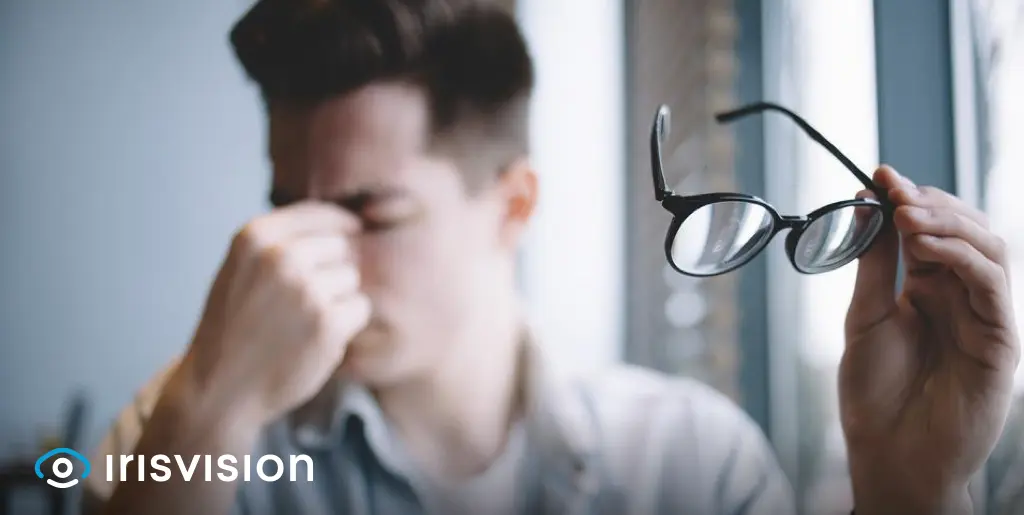
A statistical study by the Center for Disease Control and Prevention (CDC) shows that an estimated 11 million people in the US over the age of 40 have some form of vision impairment.
You may wonder, what accounts for this overwhelming figure?
Let’s find out what the facts and figures have to say.
As per the research findings of the CDC, a majority of 8 million people get defective vision due to uncorrected refractive error, while 3 million others have hereditary eye diseases, or get accidental injuries that lead to permanent vision damage.
However, despite the visually impairing conditions, most people with eye ailments do not lose their vision completely. Rather, they are left with reduced and deteriorated visual ability – referred to as low vision – for the rest of their lives.
Before we dive deeper into the causes of vision loss, let’s take a glance at what low vision looks like.
Having low vision implies that a person has a visual acuity of 20/70 or worse (after correction) in the better-seeing eye. It cannot be corrected with glasses, contacts, medicine, or surgery.
In the US, low vision affects approximately 1 in 28 people who are 40 years of age or older.
Low vision is caused by a multitude of eye problems, and the nature of vision damage depends on the disease that is causing it. Forms of low vision may include
According to the World Health Organization, low vision is classified into different categories based on its severity. A person’s visual acuity, the field of vision, and light perception all help gauge the degree of low vision.
It presents as one or more of the following conditions:
Now, let’s look at some of its most common causes. Listed below are the top five most common eye diseases that lead to low vision.
Cataracts is a common condition in people older than 40. It affects the lens of the eye.
In a healthy eye, the lens is translucent and forms a clear image of the object in front of you by focusing light that passes through it.
But in a person with cataracts, the lens becomes clouded with the remains of broken-down eye tissue, and the image it forms is blurry and unclear.

Having cataracts is similar to looking through a foggy window and seeing a smudged view of the outside. The colors appear blotched and mixed with one another, which makes it difficult to make out what you see.
ataracts begin with mild visual difficulty, but it is a progressive disease and worsens over time. The symptoms are usually noticeable until 60 years of age.
Its symptoms include:
The most common cause of cataracts is aging. According to the medical center of Ophthalmology Physicians and Surgeons, over half of Americans will have cataracts by the age of 75.
Other risk factors such as diabetes, smoking, excessive alcohol consumption, high blood pressure, and eye injuries may increase your chances of getting it.
In its early stages, ophthalmologists recommend wearing prescription glasses for vision correction. However, when the entire eye lens becomes clouded and vision deteriorates significantly enough to hinder daily life activities, surgery is done to replace the damaged lens with a new one.
Diabetic retinopathy is a condition that occurs as a result of diabetes (both type-1 and type-2). The unstable blood sugar in diabetic patients causes damage to the blood vessels within the light-sensitive tissue of the eye (retina), thus causing vision loss.
Diabetic retinopathy progresses in four stages, beginning with mild nonproliferative retinopathy and ending with severe proliferative retinopathy. Read more about it here.
It takes several years to advance to the stage of serious vision loss, however, if left untreated it can cause blindness. Research shows that diabetic retinopathy is the leading cause of blindness in American adults.
Vision changes are noticeable after a few years of onset. If you have diabetic retinopathy, you will see small black blotches spread across your field of vision, while the rest of the things appear blurry and out-of-focus.
Typical symptoms of diabetic retinopathy include:
The prime risk factor for diabetic retinopathy is diabetes. People who have had diabetes for a long time and who have poor control over their blood sugar levels are at a greater risk of developing this condition.
The blood vessels within the retina become clogged due to high blood sugar, which causes the eye to promote the growth of new ones for blood supply.
The new blood vessels are fragile and do not develop properly, so they often leak fluid and blood, causing further damage to other structures within the eye.
Diabetic retinopathy can not always be prevented, but it can be managed by adopting healthier lifestyle changes. Other treatments include anti-VEGF therapy, laser surgery, and low-vision aids, such as IrisVision, for improving eyesight. An ophthalmologist can guide you with the treatment that suits you best.
Age-related Macular degeneration (AMD) is an eye condition in which the ‘macula’ of the eye – a structure within the light-sensitive eye tissue – wears down. Deterioration of the macula leads to the loss of central vision, while peripheral vision remains intact.
1- Dry Age-related Macular Degeneration
2- Wet Age-related Macular Degeneration
Both types of AMD cause damage to the central vision. Read here to learn more about Dry and Wet AMD and how to prevent them.

In the early stages of macular degeneration, vision starts to become hazy and unclear. As opposed to the fine quality and color of images in normal vision, AMD causes reduced visual acuity in one or both eyes, followed by dark spots forming in the central visual field.
Macular degeneration usually begins as the dry form of the disease, and its symptoms develop gradually. These include:
Dry macular degeneration can turn into the wet form, which is more severe. Approximately 10% of all AMD patients develop the wet form.
If Dry AMD is not diagnosed and treated in time, it converts into Wet form. The onset of Wet AMD is sudden and worsens rapidly.
The causes of macular degeneration are most often genetic and age-related, thus the name Age-related Macular Degeneration. People aged 55 or older have a higher risk of getting AMD, especially if they have a family history of the disease.
While you cannot completely prevent AMD from occurring, you can minimize your chances of getting it by taking these preventive measures. Read this detailed guide on AMD to learn more.
Retinitis pigmentosa is a group of genetic disorders that cause damage to the retina – which is the light-sensitive tissue of the eye. It is a rare disease that causes progressive damage to your vision.
When the rod and cone cells of the retina get damaged, both color vision and night vision become compromised.

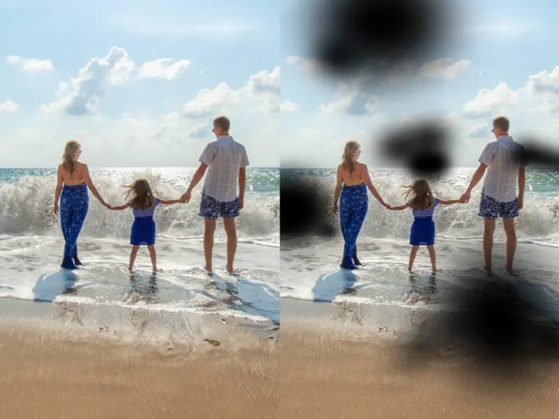
In individuals who have retinitis pigmentosa, vision appears dull and vague, and it becomes difficult to see in the dark. As the rod and cone cells undergo damage, your vision becomes restricted to the center only.
It is as if you are seeing through the end of a tunnel. Things in your peripheral view appear blacked out, while the remaining central vision looks distant and blurry.
Despite the gradual loss of vision, most people do not lose their sight completely.
The typical symptoms of retinitis pigmentosa include:
Overall visual capacity becomes poor in retinitis pigmentosa, such that you cannot carry out tasks such as driving, reading, cutting, or other activities that require precision.
A restricted field of view may even cause you trouble walking through any space without bumping into objects.
Retinitis pigmentosa is a genetic condition that is caused by the mutation of multiple genes. There are over 100 genes that contain the code for building proteins needed by cells of the retina. An abnormality in any of the genes’ structures causes damage to retinal cells and leads to retinitis pigmentosa.
Retinitis pigmentosa can be passed from parent to child and cannot be prevented. Read here to learn more about its pattern of inheritance.
For prevention of retinitis pigmentosa, you can opt for “genetic testing” to check if you may be carrying it and to determine its course and severity ahead of time.
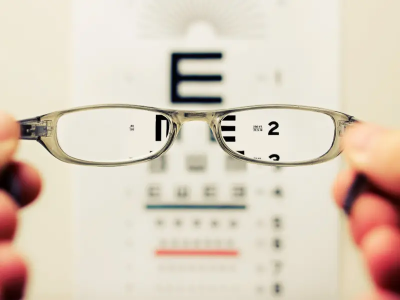
Refractive errors mean having blurred and unclear vision. They occur when the shape of the eye lens changes in a way that it cannot focus light on the retina correctly, thus showing us faulty images.
Refractive errors are the most common type of vision problem and account for over 80% of all vision impairment in the US.
We will briefly explore the most common types of refractive errors and what causes them. The four most common types of refractive errors are:
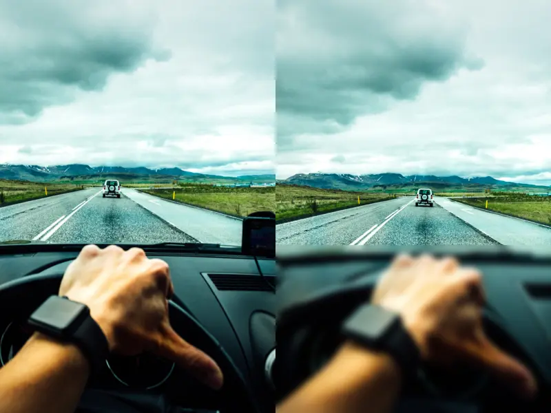
In myopia, near objects can be seen clearly while distant objects appear blurry. Myopia, or nearsightedness, often runs in families and worsens during childhood and adolescence.
If you have myopia, you will experience some obvious signs, such as:
You can correct this condition with the use of contact lenses, powered glasses, or refractive surgery.
In hyperopia, distant objects can be seen clearly while near objects appear blurry. Like myopia, hyperopia is also an inherited condition and is usually present at birth, but with healthy diet and lifestyle choices, eyesight can be improved in adulthood.
It limits your ability to focus, and you may only be able to see objects that are at a very far away distance. You may experience the following symptoms:
The corrective treatment for hyperopia is the same as that for myopia. An optometrist will give you the correct powered glasses or contact lenses for better vision. Refractive surgery is also done, usually for people whose refractive error (glass power) has become stable, typically after 18-21 years of age.
In astigmatism, both near and distant objects appear blurry and unclear. It occurs when either the lens or the cornea of the eye, which usually has a round curve, develops a mismatched curve.
This asymmetric curvature of the cornea or lens makes vision appear as if you are looking through a distorted, wavy mirror. This distortion in an image may be more in one direction, i.e. vertical, horizontal, or sideways.

Some of the symptoms of astigmatism include:
Contrary to a popular myth, astigmatism is not caused by reading in poor light, nor by using a screen too close to your eyes. Instead, an individual may have it by birth, or sometimes it can occur as a result of eye trauma or surgery.
Presbyopia is the gradual loss of the ability to focus on nearby objects. It is part of the aging process and usually starts after 40.
The lens of the eye is a flexible organ, it can contract and relax to focus on objects placed at varying distances. The lens contracts to focus on a nearby thing and produces a clear image of it.

But with age, eye muscles become weak and cannot contract, which is why we may get a blurred vision of an object placed close to us.
Some of its noticeable symptoms include:
As the saying goes, prevention is better than cure. All eye health experts encourage you to get an annual eye exam, especially after the age of 40. A regular check-up helps detect your vulnerability to developing any eye complications so they can be treated timely.
If you experience symptoms of any of the above low vision conditions, consult an ophthalmologist. Various treatments for these conditions include oral medication, corrective glasses, laser surgery, and multiple low vision aids that help you see better.
Electronic low vision aids, such as IrisVision Inspire, are among the leading visual aids that have helped restore sight for thousands of visually impaired people.
These electronic glasses allow you to navigate the world through a customised lens that is tailored to your specific needs; for example, you can magnify any object by 10x, see a virtually wider field of view, read comfortably in a special reading mode, watch videos on YouTube, and stay connected to the world around you.
Visual aids such as this offer multiple functions that help the low vision community stay in touch with their surroundings and be more independent in living their lives.
Support
See and Connect Today!
IrisVision Global, Inc.
5994 W. Las Positas Blvd, Suite 101
Pleasanton, CA 94588
Email: [email protected]
Support: +1 855 207 6665
Support
See and Connect Today!
IrisVision Global, Inc.
5994 W. Las Positas Blvd, Suite 101
Pleasanton, CA 94588
USA Email: [email protected]
Support: +1 855 207 6665
Support
See and Connect Today!
IrisVision Global, Inc.
5994 W. Las Positas Blvd, Suite 101
Pleasanton, CA 94588
Email: [email protected]
Support: +1 855 207 6665